
FIG. 5.
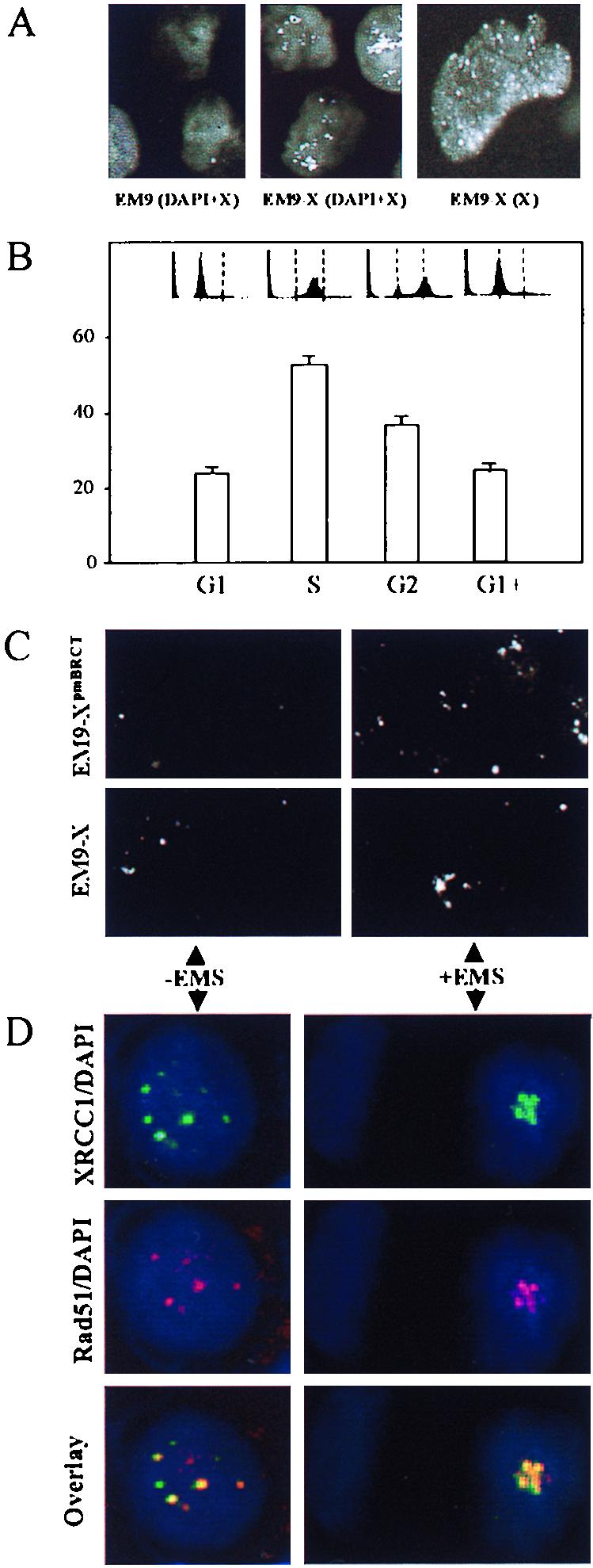
XRCC1 foci. (A) EM9 (left panel) and EM9-X (middle panel) cells immunostained with anti-XRCC1 monoclonal antibody and DAPI. White spots are XRCC1 foci, and grey background is DAPI-stained nuclear DNA. The right panel shows EM9-X cells immunostained with anti-XRCC1 monoclonal antibody. White spots are foci, and grey background is diffuse or speckled staining. (B) Frequency of XRCC1 focus-positive EM9-X cells following synchronization in G1, during transit through S phase or G2, or after maintenance in G1 throughout the experiment (G1+). The flow cytometry profiles are shown with dotted lines indicating G1 and G2. (C) Asynchronous EM9-X or EM9-XpmBRCT cells immunostained for XRCC1 (white dots) before or 8 h after EMS treatment (2 mg/ml, 1 h). Each panel depicts approximately six cells. (D) Asynchronous HeLa cells were harvested and fixed before (−EMS) or 8 h after (+EMS) EMS treatment (2 mg/ml, 1 h) and stained with DAPI plus anti-XRCC1 monoclonal antibody (green; top panels), DAPI plus anti-Rad51 polyclonal antibody (red; middle panels), or DAPI plus anti-XRCC1 and anti-Rad51 (bottom panels, overlapping antibody signals in yellow).