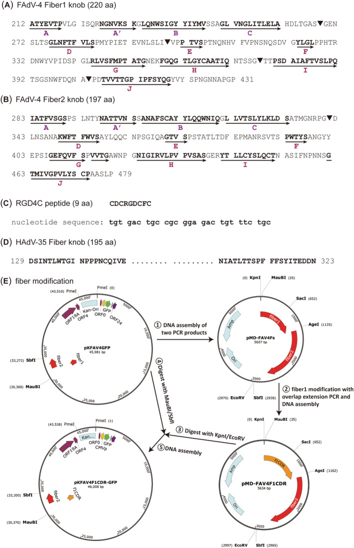
FIGURE 1.

Schematic diagram of fiber modification sites, elements and procedure. (A) Amino acid sequence of the FAdV‐4 fiber1 knob. The β‐strands, which are shown as bold letters and denoted with arrows, were predicted according to the alignment of FAdV fiber knob domains and the crystal structures of FAdV‐1 fiber1 (PDB ID 2IUN) and fiber2 (PDB ID 2VTW). 38 The sites for RGD4C insertion are labelled with a solid inverted triangle “▼”. (B) Amino acid sequence of FAdV‐4 fiber2 knob. The site for RGD4C insertion is labelled with a solid inverted triangle “▼”; and the whole knob is replaced with that of HAdV‐35 fiber in another construction. (C) Amino acid and coding sequence of RGD4C peptide. (D) Amino acid sequence of HAdV‐35 fiber knob. The sequence in the middle is abbreviated; for full annotation, please refer to the GenBank sequence of AC_000019. (E) Procedure for modification of FAdV‐4 fiber1 knob. The genome of FAdV‐4 in pKFAV4GFP originated from FAdV‐4 isolate NIVD2 (GenBank MG547384) 32
