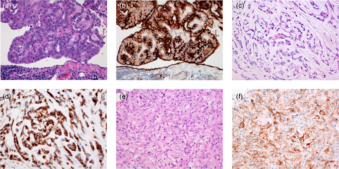
Figure 1.

Epithelioid mesothelioma with tumor cells growing in papillary pattern (a) and showing strong membranous HEG1 staining (b). Reactive mesothelial cells also show membranous or cytoplasmic HEG1 staining. Epithelioid mesothelioma with tumor cells growing in trabeculae (c). Mesothelioma cells show strong cytoplasmic HEG1 staining (d). Pleomorphic mesothelioma (e) with tumor cells showing granular cytoplasmic HEG1 staining (f)
