Figure 4.

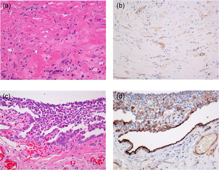
Fibrous pleuritis (a) with spindle cells showing weak cytoplasmic HEG1 staining (b). Pleural reactive mesothelial proliferation associated with pneumothorax (c) showing strong apical HEG1 staining in reactive mesothelial cells (d)

Fibrous pleuritis (a) with spindle cells showing weak cytoplasmic HEG1 staining (b). Pleural reactive mesothelial proliferation associated with pneumothorax (c) showing strong apical HEG1 staining in reactive mesothelial cells (d)