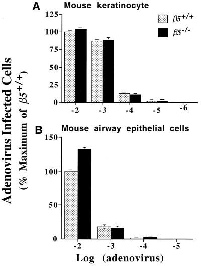
FIG. 4.
Keratinocytes (A) and airway epithelial cells (B) from wild-type and β5−/− mice were infected with a range of concentrations of adenovirus H5.010CMVlacZ; 40 h after infection, cells were treated with X-Gal and blue-stained cells were counted. The results are expressed as a percentage of the number of positive wild-type cells after incubation with the highest concentration of adenovirus used. Results are the mean (± standard error of the mean) of three experiments.

