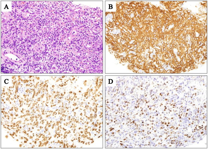
Fig. 1.
Histological findings of a patient with DLBCLOIIA (Patient 13). (A) Diffuse infiltration of lymphoma cells (hematoxylin and eosin staining; original magnification, ×1000). (B) lymphoma cells were positive for CD20 (original magnification, ×1000). (C) Ki-67 was expressed in approximately 60% of the lymphoma cells (original magnification, ×1000). (D) MYC was expressed in approximately 60% of the lymphoma cells (original magnification, ×1000).

