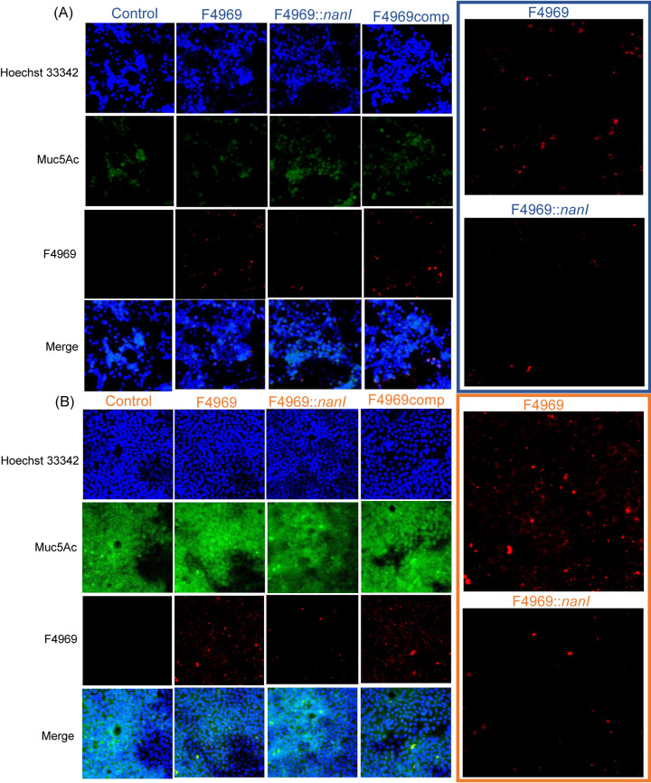
FIG 7.
Microscopic comparison of mucin production by the HT29 and MTX-E12 cell lines, as well as detection of C. perfringens attachment to HT29 (A) and MTX-E12 (B) cells. Attachment of C. perfringens F4969, its nanI null mutant, and complementing strain to HT29 or MTX-E12 cell lines as detected by Olympus confocal laser scanning biological microscope (FluoView FV1000) with FV10-ASW (version 1.4) software. All pictures were taken at a magnification of ×400. Red, C. perfringens; green, Muc5Ac; blue, mammalian cell nuclei. Panels at right show enlarged view of attached wild-type F4969 and nanI null mutant in panels at left.

