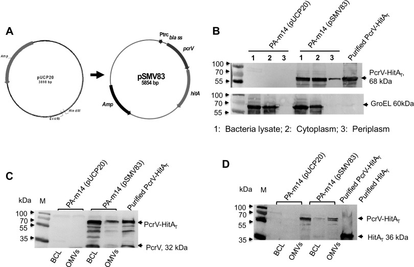
FIG 2.
Enhancement of the PcrV-HitAT fusion antigen in P. aeruginosa OMVs. (A) Construction of the pSMV83 plasmid, containing a fusion gene encoding the PH fusion antigen. The Ptrc-bla ss-pcrV-hitAT gene fragment was inserted into pUCP20. (B) Comparison of PH amounts in different cell fractions. The total-cell lysates and subcellular fractions, including the cytoplasmic and periplasmic fractions, were prepared from the PA-m14(pUCP20) and PA-m14(pSMV83) strains. The cells were grown in LB broth at 37°C for 16 h, as described in the Materials and Methods section of the supplemental material. Fractions with 25-μl volumes from cultures grown to an OD600 of 0.8 were evaluated by immunoblotting with a PcrV-specific polyclonal mouse antibody. GroEL was used as a cytoplasmic marker for fractionation. Five micrograms of purified PH protein was used as a loading control. (C) The PH fusion antigen in the BCL (6 × 10⁸ CFU bacterial lysate) and 33 μg OMVs from wild-type PA103 or mutant PA-m14 was detected by Western blotting using mouse primary anti-PcrV antibodies. PH protein (3.5 μg) was used as a loading control. (D) The PH fusion antigen in the BCL (6 × 10⁸ CFU bacterial lysate) and 33 μg OMVs from wild-type PA103 or the PA-m14 mutant was detected by Western blotting using mouse primary anti-HitAT antibodies. PH protein (3.5 μg) was used as a loading control.

