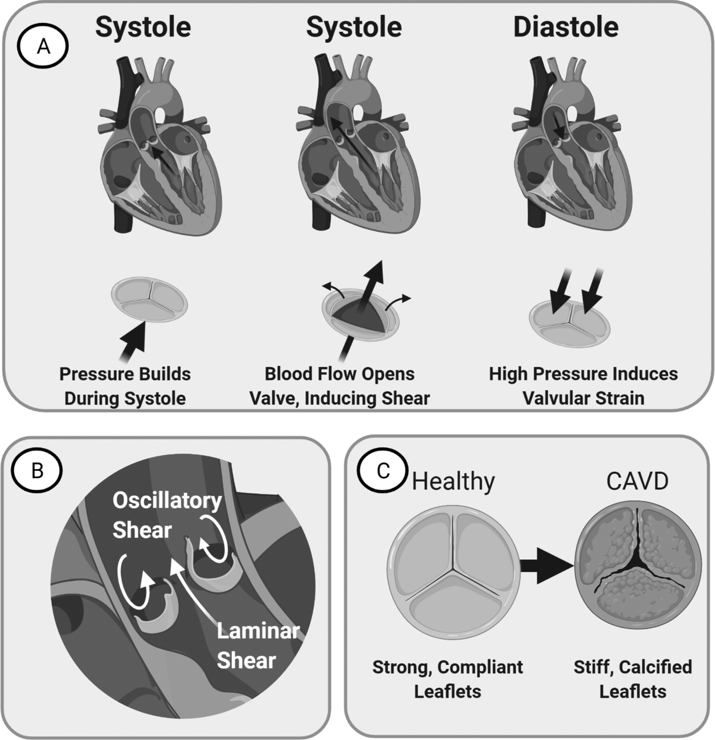
Figure 1. Anatomy and Physiology of the Aortic Valve in homeostasis and disease.

(A) Left: Valve just before systole. Blood flow assists opens the valve. Middle: Valve during Systole. Leaflets ends bend to create an opening for blood flow. Right: Valve during Diastole. Valve provides seal that resists back-flow pressure, inducing strain. (B) Image of the valve demonstrating laminar flow on the ventricularis side and oscillatory flow on the fibrosa side. (C) CAVD manifests as valvular thickening with stiffening and calcification that reduces leaflet mobility over time.
