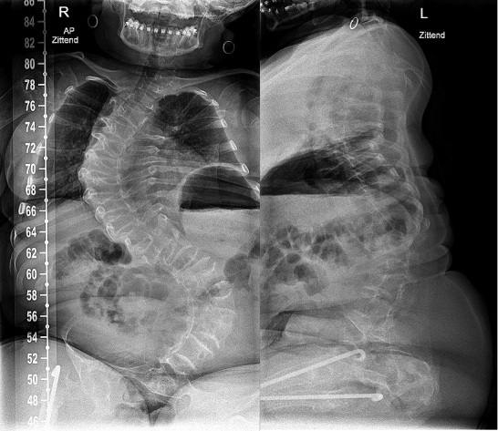
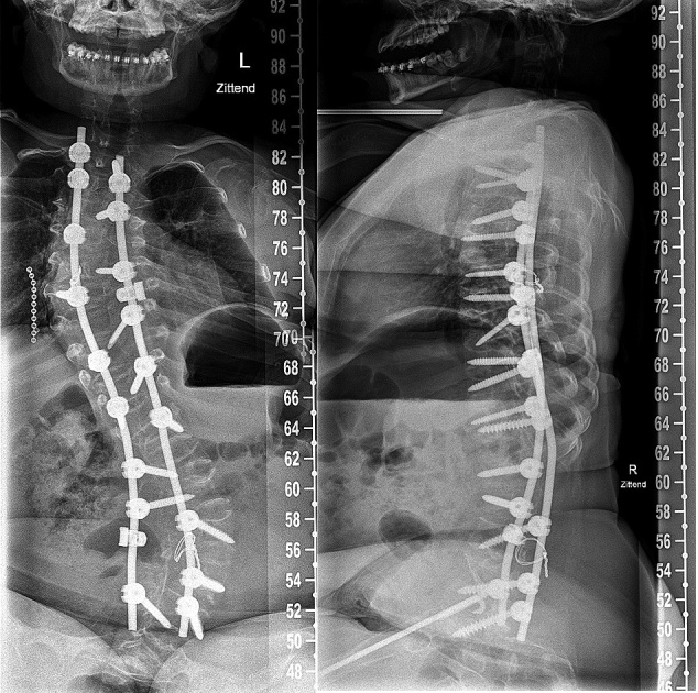
Figure 6.
a. Preoperative radiograph of a 15-year-old patient with OI type III with progressive scoliosis.
b. Note the decreased space for pedicle screws in the codfish vertebrae and increased diameter of the intervertebral disks. Augmentation with a sublaminar wire at level L4.


