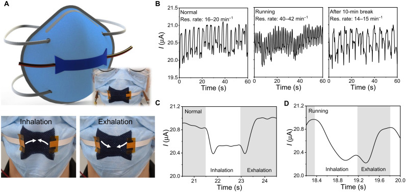
Fig. 4. oCVD PEDOT on the mask for respiratory rate sensor.
(A) Schematic and photo images of oCVD PEDOT sensor fabricated on a disposable mask. The top images demonstrate a schematic of the mask sensor and an actual sensor image; the bottom photo images represent the instant deformation of the mask during inhalation and exhalation. (B) Breath patterns measured at an initial state (left), after 10-min exercise (middle), and further 10-min break (right). A single respiratory pattern, measured at (C) the normal and (D) as soon as after the exercise. Photo credit: Michael Clevenger and Hyeonghun Kim, Purdue University.

