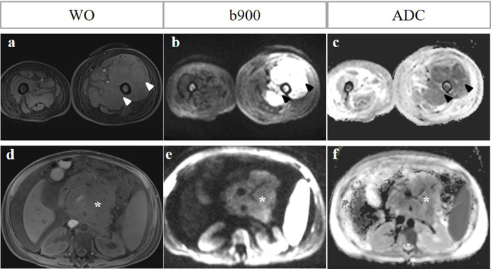
Figure 4.
Paramedullary versus extramedullary disease. A 51-year-old female with relapsed MM post-haematopoetic stem cell autograft. WO Dixon, b900 DWI and ADC map show paramedullary disease in the left mid femoral shaft with a large paramedullary mass (arrowheads, a-c). Note also extramedullary disease in the form of a retroperitoneal nodal mass and pancreatic involvement with associated ascites (asterisk, d–f). The signal intensity of the retroperitoneal mass of the b900 image is lower than expected as the patient had already commenced systemic therapy 7 days before the MRI.

