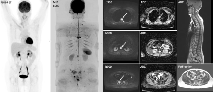
Figure 7.
A 65-year-old female with history of right invasive lobular breast carcinoma presenting with raised tumour marker CA15-345 U/ml and back pain. The tumour showed lymphovascular invasion; with ER8 PR0 Her-2 negative. WB-MRI show multiple vertebral and pelvic bone metastases that were occult on PET-CT without FDG tracer uptake.

