
Figure 1.
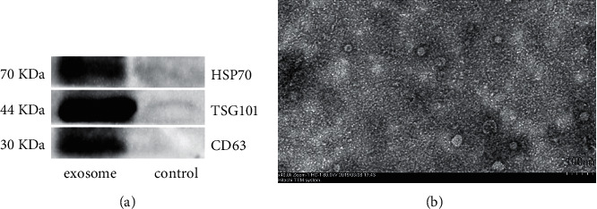
Identification of exosomes derived from RM-1. (a) Biomarkers CD63, HSP70, and TSG101 expression in exosomes detected by Western blot. Exosomes-depleted supernatant was set as the control group. (b) Purified exosomes observed by electron microscopy after ultrafiltration centrifugation; result showed exosomal pallet-like lipid bilayer vesicles and the average diameter was among 30–80 nm. The scale bar is 100 nm.