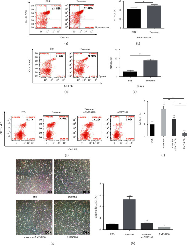
Figure 3.

Exosomes induced MDSCs expansion and migration. The recruitment could be inhibited by AMD3100 both in vivo and in vitro. After injecting exosomes to normal mice via the tail vein 3 times per week for 2 weeks, the percentage of MDSCs in the BM ((a), (b)) and spleen ((c), (d)) was examined by flow cytometry. ((e), (f)) The percentage of MDSCs in tumor tissue of PBS, exosomes, exosomes plus AMD3100, and AMD3100 treatment groups measured by flow cytometry, respectively. ((g), (h)) In transwell chemotaxis assay, MDSCs from BM of mice bearing tumors were divided into PBS, exosomes, exosomes plus AMD3100, and AMD3100 treatment groups in the upper chamber; after cocultured with CXCL12 for 24 h, cells were counted and averaged through the selection of 5 random fields/well under a light microscope to detect the chemotaxis effect of CXCL12 to MDSCs. ∗P < 0.05 and ∗∗P < 0.01 compared with the PBS group.
