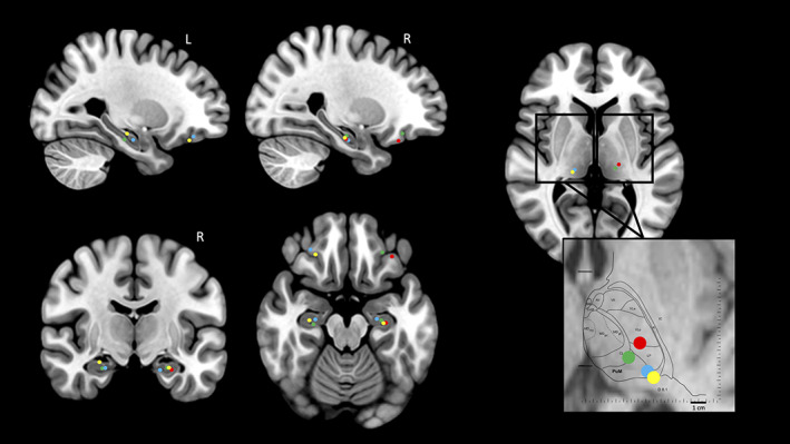
FIGURE 1.

Recording contact locations represented on MNI brain template and Morel's thalamic atlas. The reconstruction of hippocampus, orbito‐frontal cortex, and thalamus contacts locations on the MNI brain template is presented for the four patients. For the thalamus, left and right side are merged in the zoom window which shows the location of the different nuclei on the Morel's atlas. R, right; L, left. Each color refers to a patient: Patient 1: blue circle; patient 2: red circle; patient 3: green circle; patient 4: yellow circle. Thalamic nuclei: PuM, medial pulvinar; CL, central lateral; L, lateral posterior; VLp, ventral lateral posterior. The two horizontal lines indicates the positions of the anterior and posterior commissures. D8.1: horizontal plane 8.1 mm dorsal to the horizontal inter‐commissural plane
