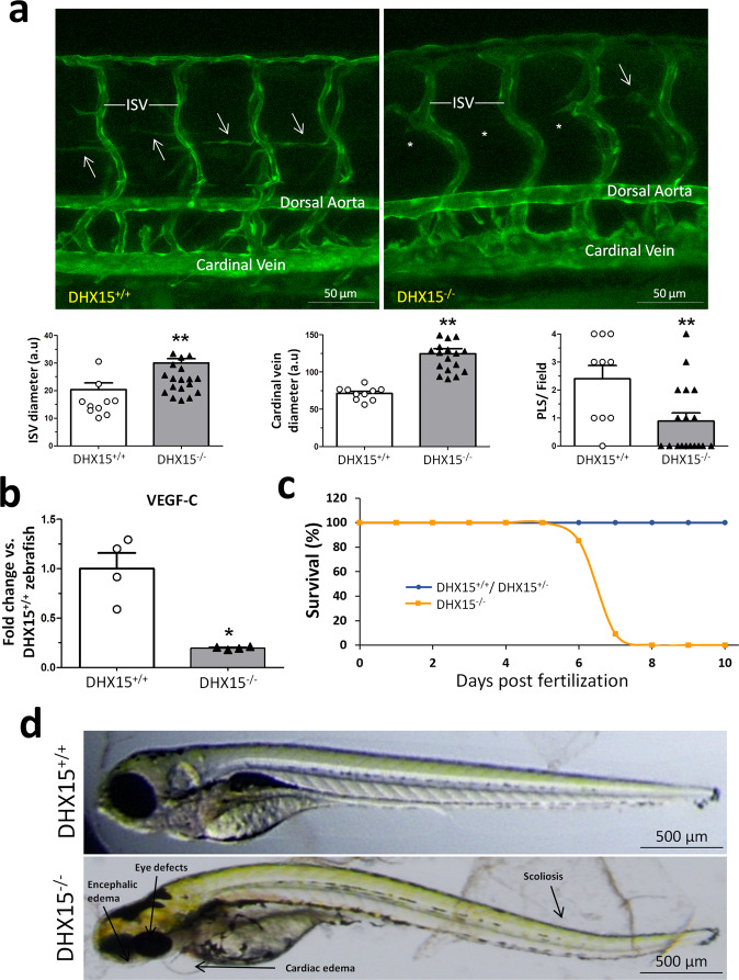
Fig. 3. Characterization of embryonic vascular anomalies associated with DHX15 gene deficient in zebrafish.
a Representative vascular images of DHX15+/+ and DHX15−/− larvae at 5 day post fertilization (dpf) revealing a reduced formation of the parachordal line (arrows). Asterisks denote the absence of these vascular structures in DHX15−/− animals. Quantifications of cardinal vein diameter, intersegmental vessels (ISV), and number of parachordal lymphangioblast strings (PLS) are shown in the graphs. **p < 0.01 vs. wild-type zebrafish, unpaired two-tailed Student’s t-test (n = 10 and n = 18 for the DHX15+/+ and DHX15−/− conditions, respectively). Arbitrary units: a.u. b RNA extraction of zebrafish embryos at 4 dpf from either wild-type or DHX15 knockout larvae was performed. mRNA expression was analyzed by RT-qPCR. The graph shows the different expression levels of the VEGF-C gene in the DHX15+/+ and DHX15−/− conditions. mRNA levels are shown as fold change relative to HPRT mRNA levels. *p < 0.05 vs. wild-type unpaired two-tailed Student’s t-test (n = 4 biologically independent samples for each condition). c Survival assessment assay. The graph shows the larvae survival rate through the first 10 dpf according to their different genotype (n = 15 zebrafish larvae for each condition). d Representative images comparing wild-type and DHX15−/− larvae at 7 dpf where morphological defects including encephalic and cardiac edema, scoliosis, and impaired neural/eye growth are evident. All bar graphs are presented as mean ± SEM.

