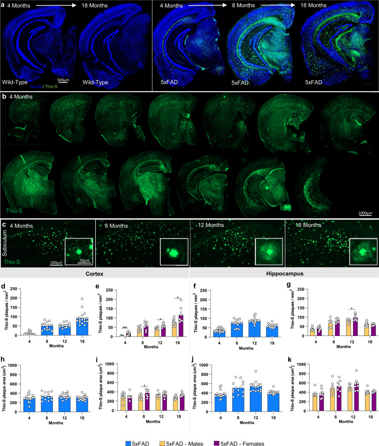
Fig. 4. Fibrillar amyloid plaques increase in size and number in 5xFAD aged mice.
5xFAD plaque burden was assessed with Thio-S staining at each time point. (a,b) Representative stitched brain hemispheres of 5xFAD shown with Thio-S staining at the 4- and 18-month and 4, 8, and 18 mo timepoints respectively, counter stained for NeuN. (b) Representative stitched whole brain hemispheres of 5xFAD (rostral to caudal) shown with Thio-S staining at the 4 month timepoint. (c) Representative images of plaques in 5xFAD mice across timepoints displaying a “halo” effect at 12 and 18 months. (d–g) Quantification for number of Thio-S positive plaques in the cortex and hippocampus by genotype and sex. (h–k) Quantification of average plaque area in the cortex and hippocampus by genotype and sex. Data are represented as mean ± SEM. *P ≤ 0.05, **P ≤ 0.01, ***P ≤ 0.001, ****P ≤ 0.0001, n = 6 per sex per age.

