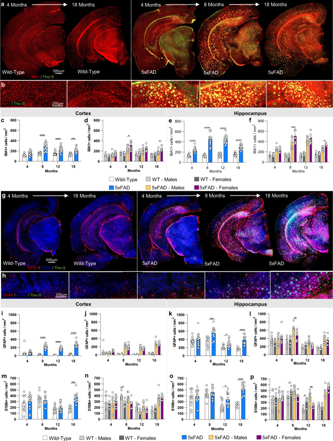
Fig. 6.
Immunostaining of microglia and astrocytes. Brains of mice at each timepoint were sliced and immunostained for IBA1, GFAP and S100ß to reveal any changes in microglial, astrocytic. (a,b) Representative stitched brain hemispheres of WT and 5xFAD shown with IBA1/Thio-S staining at the 4- and 18-month and 4, 8, and 18 months timepoints, respectively. (c–f) IBA1 immunostaining for microglia reveals both age-related changes in WT and 5xFAD microglial number, and differences between genotypes in cortex and hippocampus. (g,h) Representative stitched brain hemispheres of WT and 5xFAD shown with GFAP/ S100ß/Thio-S staining at the 4- and 18-month and 4, 8, and 18 months timepoints, respectively. (i–p) Astrocyte number is assessed via GFAP (i-l)) and S100ß staining (m–p) in the cortex and hippocampus. Data are represented as mean ± SEM. *P ≤ 0.05, **P ≤ 0.01, ***P ≤ 0.001, ****P ≤ 0.0001, n = 6 per group.

