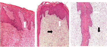
Figure 6. Post-treatment biopsies showing a decreased epidermal proliferation and an increased fibroplasia of connective tissue core (blunt finger-like projection) (white arrow). The dermal layer was infiltrated with lymphocytic cells (black arrow) (H&E stain).

