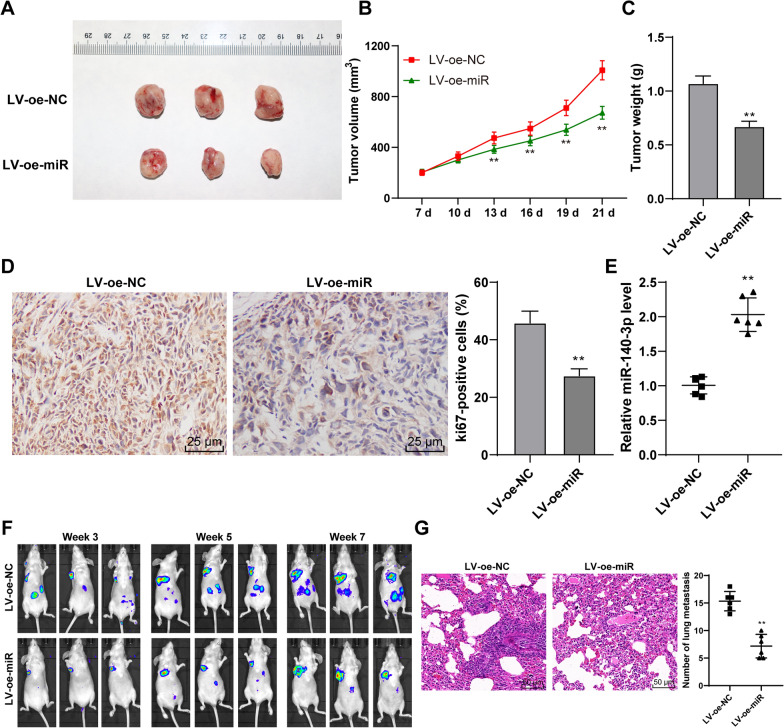
Fig. 3.
Overexpression of miR-140-3p inhibited the growth and metastasis of GC. Nude-mouse transplanted tumor model was established using AGS cells with overexpression of miR-140-3p. A Representative image of a transplanted tumor model. B The volume of the tumor during modeling. C The weight of the tumor on day 21. D The positive rate of ki67 in GC tissues was measured by immunocytochemistry. E The expression of miR-140-3p in tumor tissues was detected by RT-qPCR. Nude-mice lung metastasis models were established using AGS cells with overexpression of miR-140-3p. F Metastatic area of GC was detected using an in vivo imaging system. G The number of pulmonary metastasis was observed by HE staining. Data in B, C, D were presented as mean ± standard deviation. Comparison among panels in C, D, F, G was performed using one-way ANOVA. Comparison of data in B was performed using two-way ANOVA, followed by Tukey's multiple comparisons test. **p < 0.01. LV-oe-miR: The lentiviral overexpression vector of miR-140-3p. LV-oe-NC: negative control of lentiviral overexpression vector

