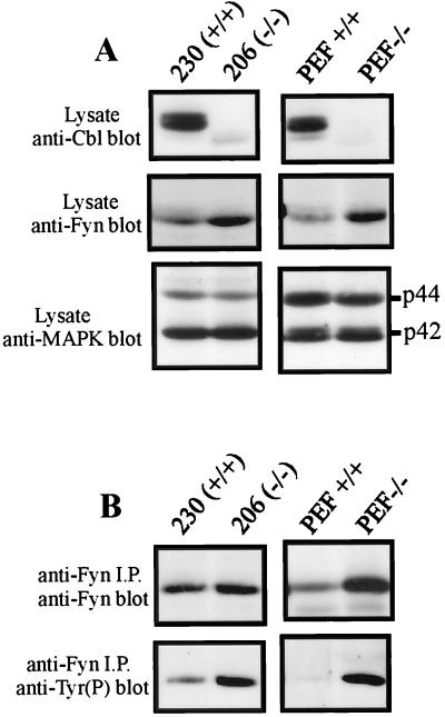
FIG. 7.
Fyn phosphorylation and protein levels are increased in Cbl−/− cell lines. (A) Total-cell lysates from the Cbl−/− T lymphoma cell line 206 and control line 230 (150 μg) were separated by SDS-PAGE and transferred to a PVDF membrane. Similarly, total cellular protein (200 μg) from the Cbl−/− fibroblast line PEF−/− or the control line PEF+/+ was immunoblotted with anti-Cbl (top panel), anti-Fyn (middle panel), or anti-MAP kinase (MAPK) (bottom panel) antibody. (B) Fyn protein was immunoprecipitated (i.p.) from T-cell or fibroblast lysates, resolved by SDS-PAGE, and transferred to a PVDF membrane. The membrane was then probed with anti-Fyn antibody, stripped, and reprobed with antiphosphotyrosine [Tyr(P)] antibody.

