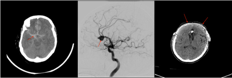
Figure 1. Imaging related to the clinical course of patient 1. (A) Head CT obtained on presentation demonstrated SAH secondary to a ruptured right pericallosal aneurysm. (B) Angiogram obtained after presentation demonstrating a large right pericallosal aneurysm. (C) Post-operative head CT after bilateral DCHC for refractory elevated ICP.
SAH, subarachnoid hemorrhage; DCHC, decompressive hemicraniectomy; ICP, intracranial pressure

