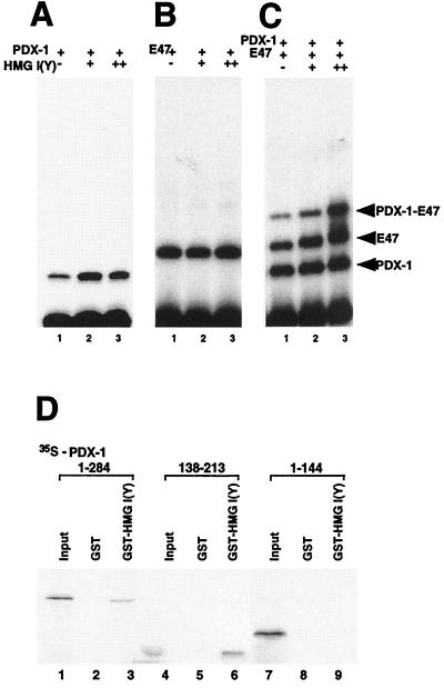
FIG. 9.
HMG I(Y) interacts with the activation complex on the E2A3/4 minienhancer. (A) The homeodomain of PDX-1 (160 pg) was incubated with the 32P-labeled rat insulin E2A3/4 probe with GST-HMG I(Y) (160 ng in lane 2; 320 ng in lane 3) or GST alone (320 ng in lane 1; 160 ng in lane 2) and analyzed by EMSA. (B) The bHLH domain of E47/Pan1 (6 ng) was incubated with the 32P-labeled rat insulin E2A3/4 probe with GST-HMG I(Y) (160 ng in lane 2; 320 ng in lane 3) or GST alone (320 ng in lane 1; 160 ng in lane 2) and analyzed by EMSA. (C) The homeodomain of PDX-1 (160 pg) and bHLH domain of E47/Pan1 (6 ng) were incubated with the 32P-labeled rat insulin E2A3/4 probe with GST-HMG I(Y) (160 ng in lane 2; 320 ng in lane 3) or GST alone (320 ng in lane 1; 160 ng in lane 2) and analyzed by EMSA. (E) In vitro-translated, 35S-labeled PDX-1 protein (wild type and deletion mutants) was incubated with GST alone or with GST-HMG I(Y). Bound proteins were immobilized on glutathione-Sepharose beads and resolved by SDS-PAGE followed by autoradiography. Ten percent of the 35S-labeled PDX-1 proteins were loaded in lanes 1, 4, and 7.

