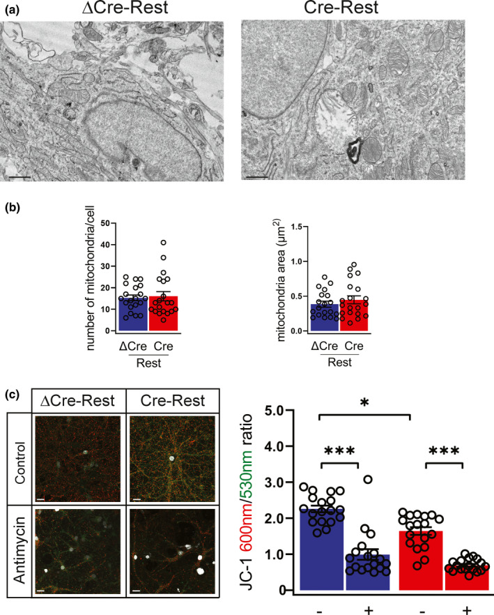
FIGURE 3.

REST‐defective neurons have impaired mitochondrial membrane potential in the absence of morphological alterations (a,b) Representative micrographs (a) and quantification (b) of the number (left panel) and area (right panel) of mitochondria in ΔCre‐ and Cre‐transduced neurons. n = 20 neurons from 3 independent preparations. Scale bar, 0.5 μm. (c) Representative images (left) and quantification (right) of JC‐1 staining in ΔCre‐REST (blue bar) and Cre‐REST (red bar) neurons in the presence or absence of antimycin (40 mM, 1 h). JC‐1 accumulates in mitochondria in a ΔΨm‐dependent manner; it exists as monomer at low concentrations (emission, 530 nm) and forms aggregates at higher concentrations (emission, 600 nm). Antimycin A (40 mM, 1 h), a complex III inhibitor, leads to a rapid breakdown of ΔΨm, confirming the dynamic assay properties. Scale bar, 5 µm. n = 18 coverslips from 4 independent preparations. Graphs show means ± sem. **p < 0.01; ***p < 0.001; Kruskal–Wallis/ Dunn's tests
