
FIGURE 1.
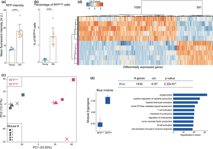
p16‐RFP expression is increased in the brain of aged p16‐3MR mice and abundantly express inflammatory and microglia genes. (a) Mean fluorescent RFP intensity of all viable cells in young compared to old brains. ****p<0,0001. (b) Percentage of viable cells positive for RFP in young mouse brains compared to old. ****p<0,0001. (c) PCA plot of bulk sequenced RFPLow and RFPHigh cells from old mouse brains. (d) Heatmap of all differentially expressed gene between the RFPLow and RFPHigh samples. E: Expression and gene‐ontology analysis of a WGCNA module enriched in RFPHigh samples