
FIGURE 1.
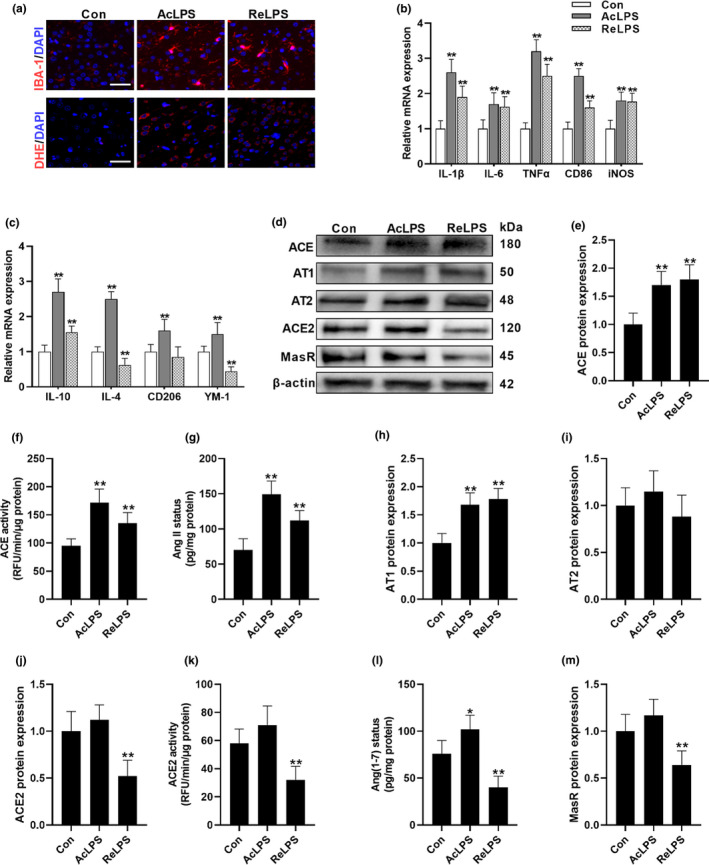
LPS exposure shifts the balance of brain RAS. (a) The impacts of acute LPS (AcLPS) or repeated LPS (ReLPS) exposure on microglial activation (IBA‐1 staining) and ROS generation (DHE staining) in the brain cortex of C57BL/6 mice. (b) Biomarkers of microglial M1 phenotype mRNA expression. (c) Biomarkers of microglial M2 phenotype mRNA expression. (d–m) Alterations of ACE/AngII/AT1 and ACE2/Ang(1–7)/MasR pathways following LPS treatment. (d) Representative western blots. ACE protein expression (e) and activity (f). (g) AngII concentration. (h) AT1 protein expression. (i) AT2 protein expression. ACE2 protein expression (j) and activity (k). (L) Ang(1–7) concentration. (m) MasR protein expression. Scale bar = 50 μm. Data are means ± SD (n = 7–9). *p < 0.05, **p < 0.01 compared to control group