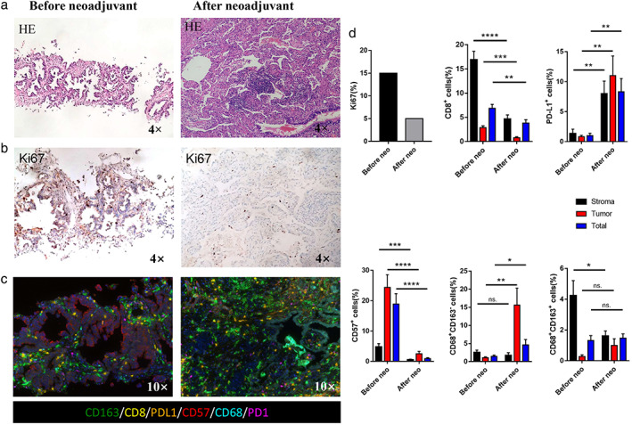
FIGURE 3.

Comprehensive pathological evaluation. (a) Hematoxylin and eosin (HE) staining. Fibrosis and lymphocyte infiltration could be seen on the surgical specimen after crizotinib treatment. (b) Ki67 staining. (c) Multiple immunohistochemistry staining on CD163, CD8, PDL1, CD57, CD68, and PD1 before and after neoadjuvant crizotinib. (d) Quantitative analysis for staining data. *p < 0.05; **p < 0.01; ***p < 0.001. Neo, neoadjuvant therapy; ns, not significant
