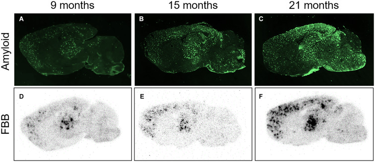
FIGURE 3.
FBB ex vivo autoradiography in comparison to amyloid plaque load. (A–C) Photographs of parasagittal brain sections from transgenic ARTE10 mice stained by immunofluorescence against Aβ plaques (antibody 6E10) at 9 (A), 15 (B), and 21 months of age (C). Plaque load is increasing with age in ARTE10 mouse brain. (D–F) Increasing plaque load is also picked up by FBB ex vivo autoradiography on adjacent brain sections of 9- (D), 15- (F), and 21-month-old ARTE10 mice (F).

