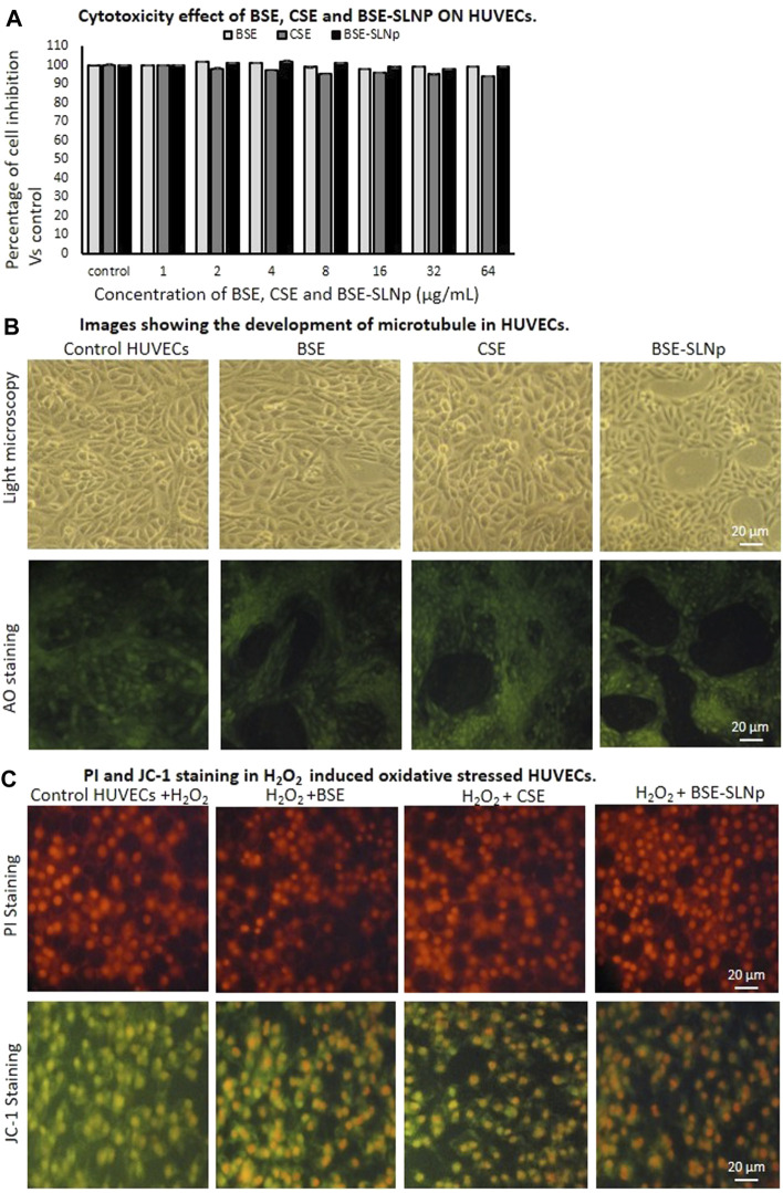
FIGURE 6.
In vitro cytotoxicity (6A), microtubule development (6B), PI and JC-1 (6C) stained microscopic image analysis of vehicle control, BSE, CSE, and BSE-SLNp treated H2O2 induced oxidative stressed HUVECs after 48 h. Each value is means ± SD (n = 6). In PI staining, the nucleus appeared to be normal and there are no signs of shrunken, pyknosis, or apoptotic nucleus. BSE-SLNp treated adipocyte showing high J-aggregates directly representing active mitochondria (high MMP, Δψm).

