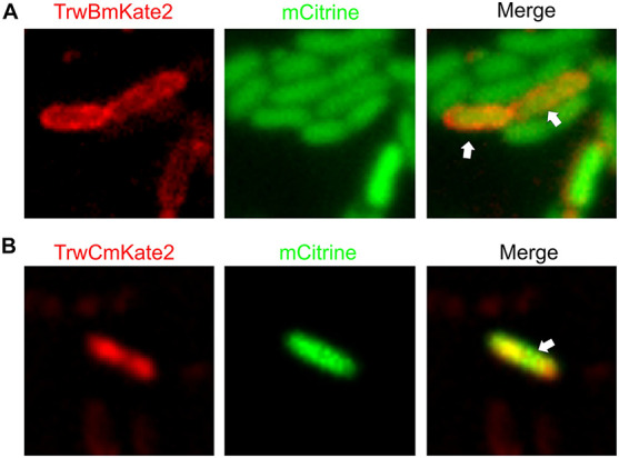
FIGURE 4.

Visualization of transconjugant cells. MG1655 donor cells hosting R388:trwBmKate2 (A) or R388:trwCmKate2 (B) were mixed with UB1637 recipient cells expressing mCitrine. Conjugation was performed in enriched M9 minimal media plates and then, cells were transferred to the microscope for image-recording. Images were recorded at different emission wavelengths (540 and 630 nm, for mCitrine and mKate2, respectively) at the same location, in order to discriminate donor, recipient and transconjugant cells. Merged images show the transconjugant cells in orange (right panel). These transconjugants are UB1637 cells expressing mCitrine that, after receiving the R388 plasmid, have started to express TrwBmKate2 (A) or TrwCmKate2 (B), encoded by the plasmid.
