FIGURE 5.

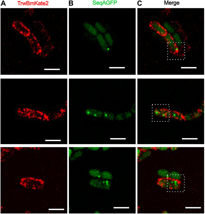
Conjugative R388 transfer visualized as fluorescent SeqA-GFP foci in recipient cells. MG1655 dam+ donor cells hosting R388:trwBmKate2 were mated with dam– recipient cells expressing SeqA-GFP from the chromosome. Conjugation was performed in enriched M9 minimal media plates and then, cells were transferred to the microscope for image-recording. Transconjugants were monitored by the presence of fluorescent foci, indicating the binding of SeqA-GFP to hemy-methylated DNA. In some transconjugant cells, up to three independent foci were detected. Images show the same pad of transconjugant cells recorded at different emission wavelengths: 630 nm for mKate2 (A, red) and 540 nm for SeqA-GFP (B, green). The merged images in panel (C) show the fluorescent foci of SeqA-GFP plus TrwBmKate2 in the membrane, which is expressed from the recently transferred R388 plasmid. (Scale bar: 2 μM).
