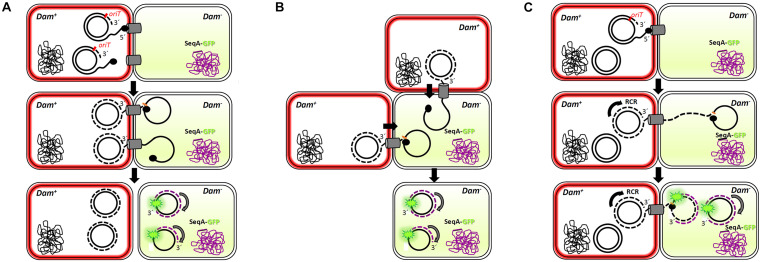
FIGURE 6.
Multi-transfer of conjugative R388 plasmid. The passage of more than one copy of the conjugative plasmid from donor to recipient cells, as shown by the presence of more than one foci in the recipient cell (Figure 5), is compatible with three models. Dam+ donor cells contain R388 plasmid copies that express TrwB-mKate2 protein, coloring the membrane in red. Dam– recipient cells express SeqA-GFP protein from the chromosome, under the control of the native SeqA promoter. Methylated DNA is represented in black, whereas no methylated DNA is in purple. SeqA-GFP expression in the recipient cell is diffuse in the absence of hemi-methylated DNA. Only when a methylated copy of the plasmid is transferred and DNA replication starts in the recipient cell, SeqA-GFP is able to form compact foci. In model 1 (A), conjugation might occur simultaneously from two or more secretion systems in contact with the same recipient cell, giving rise to at least two fluorescent foci. In model 2 (B), two donor cells would transfer the plasmid simultaneously to the same recipient cell. In model 3 (C), only one secretion system is functional but, since the transfer of plasmid ssDNA is accomplished of rolling circle replication (RCR) in the donor cell, multiple single-stranded linear copies of the plasmid (a concatemer) would be transferred bound to the relaxase TrwC. Once in the recipient cell, a new cleavage by TrwC on the nic site would re-circularize the plasmid DNA, resolving as many plasmid copies as those present in the concatemer. The synthesis of the complementary non-methylated strand would be associated to SeqA-GFP binding, observed as fluorescent foci.

