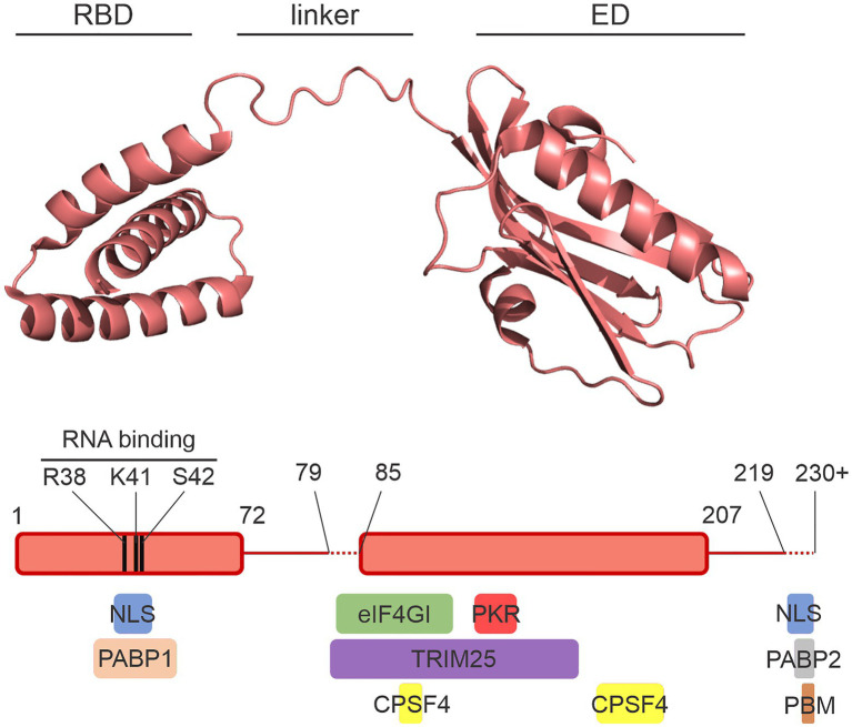
Figure 1.
The NS1 protein. A crystal structure of the NS1 protein from A/Vietnam/1196/2004 (H5N1) is shown with the RNA-binding domain (RBD), the effector domain (ED), and the flexible interdomain linker labeled. The diagram below shows the amino acid limits of the domains. The variable lengths of the linker and the structurally disordered C-terminal tail are represented with dashed lines. Colored boxes below the schematic illustrate the approximate regions of NS1 involved in interactions with a select set of host proteins. NLS, nuclear localization sequence; PBM, PDZ-binding motif. Image was prepared with PyMol using the crystal structure from Mitra et al. (2019; PDB ID: 6O01).

