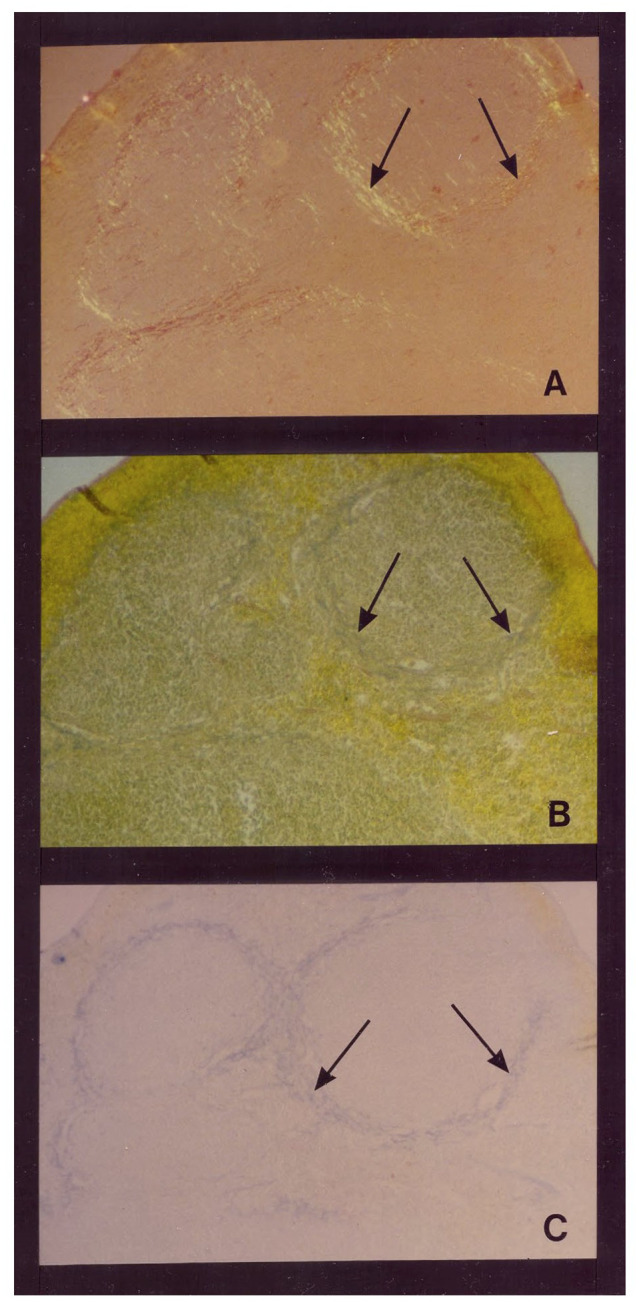
Figure 1.

Temporal relationship between AA amyloid deposition and heparan sulfate glycosaminoglycan (GAG) accumulation in the spleen in a mouse model of experimental AA amyloidosis. AA amyloid deposition and sulfated GAG deposition in the perifollicular areas of spleen at 48 h post-induction (with amyloid enhancing factor + silver nitrate (Snow, 1986). (A) Splenic amyloid (arrowheads) has extended to most perifollicular areas. Congo red staining for amyloid deposits under polarized light. (B) Positive Sulfated Alcian-Blue (SAB; arrowheads) seen in the exact perifollicular areas as amyloid deposits. (C) Positive Alcian blue staining at 0.7 M magnesium chloride demonstrating accumulation of primarily highly sulfated GAGs at amyloid deposition (Snow, 1986).
