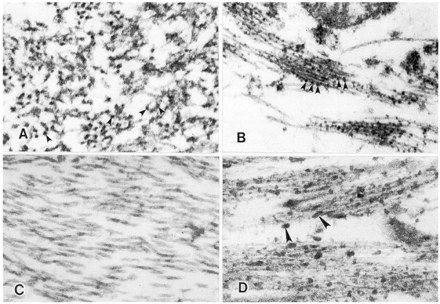
Figure 5.

Proteoglycans visualized in neuritic plaques and neurofibrillary tangles as demonstrated by Ruthenium red and Cuprolinic blue staining. (A) Ruthenium red staining of neuritic plaque in the hippocampus of a 92-year old female with AD. Positive Ruthenium red granules (arrowheads) are specifically localized along amyloid fibrils present with the neuritic plaque. (B) Ruthenium red staining of paired helical and straight filaments in a neurofibrillary tangle in the hippocampus of a 72-year old male with AD. Positive Ruthenium red granules are observed (arrowheads) specifically localized to the paired helical and straight filaments. Orig. Mag., ×80,000. (C) In the absence of Cuprolinic blue, no apparent staining of paired helical and straight filaments in a neurofibrillary tangle is observed in the hippocampus of a 92-year old female with AD. Original mag. ×80,000. (D) In the presence of Cuprolinic blue at 0.3 M magnesium chloride, proteoglycans are visualized as positive Cuprolinic blue filaments (arrowheads) in intimate association with bundles of paired helical and straight filaments in a neurofibrillary tangle in the hippocampus of a 92-year old female with AD. Original Mag. ×80,000 (Snow et al., 1989b).
