
FIGURE 1.
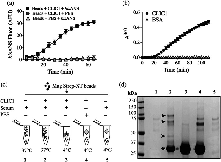
Fishing for new ECs in serum using CLIC1 as a bait protein. (a) Beads bearing bound CLIC1 were supplemented (or not) with 10 μM bisANS and then incubated at 37°C. Mean bisANS fluorescence (in arbitrary fluorescence units, AFU) ± range (n = 2) are plotted. In some cases, the error bars are too small to be visible. Result is representative of two independent experiments. (b) Amorphous aggregation of CLIC1 at 37°C measured as changes in absorbance (at 360 nm) over a period of 115 min. BSA was used as a non‐aggregating control protein. Mean absorbances (in arbitrary units, AU) at 360 nm ± SE (n = 3) are plotted. In many cases, the error bars are too small to be visible. (c) Schematic of experimental design using CLIC1 as bait to retrieve putative chaperones from human serum. Additives are indicated above each tube icon, and the temperature of incubation is indicated below the tubes. (d) Image of a completed SDS PAGE analysis of samples of bead eluates from beads treated as in (c). The numbering of lanes shown corresponds to the numbered treatments in (c). Molecular mass markers and their respective masses (kDa) are shown at the left of the image. Asterisk indicates the position of the major CLIC1 band; small arrowheads indicate the positions of bands enriched in the eluate from beads incubated with CLIC1 and serum at 37°C (compare Lane 2 with Lane 3)