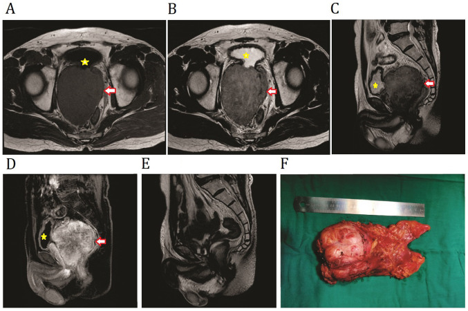
Figure 1.
Preoperative MRI imaging of primary prostatic extra-gastrointestinal stromal tumor. Non-contrast (A) T1W1 and (B) T2W1 transverse images showing a mass between the bladder and the rectum. (C) T2WI and (D) T1WI contrast-enhanced sagittal plane of the tumor was predominantly confined to the prostate. (E) Sagittal view of T2WI MRI revealed no sign of recurrence at 1 year following surgery. (F) Image of the resected sample in the present case. The tumor is indicated by the arrows and the compressed bladder is indicated by the asterisks. WI, weighted image.

