
FIGURE 6.
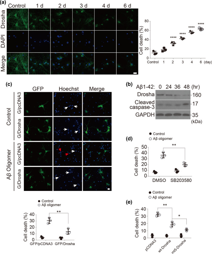
Aβ oligomers decrease Drosha levels and induce apoptosis in neurons and Drosha overexpression prevents Aβ oligomers‐induced neuronal death. (a) Primary cortical neurons at 14 DIV were treated with Aβ 1‐42 oligomers (1 μM) or its control (Aβ 42‐1) for 1–6 days. The neurons were stained with Drosha and DAPI for fluorescence microscopy analysis. Scale bar, 20 μm. Quantitative analysis of the neurons with condensed or fragmented nuclei (over 200 neurons were counted, n = 3). (b) Rat primary cortical neurons at 14 DIV were treated with Aβ 1‐42 oligomers (1 μM) for the indicated time and the levels of Drosha, cleaved caspase‐3 and GAPDH were blotted. Data showed here are the representative one from three independent experiments. (c) Primary cortical neurons at 14 DIV were co‐transfected with GFP/pcDNA3 or GFP/Drosha for 12 h and treated with Aβ 1‐42 oligomers (1 μM) or its control (Aβ 42‐1) for 48 h. The neurons were stained with Hoechst and fixed with paraformaldehyde for fluorescence microscopy analysis. The white arrows indicate the nucleus of neurons transfected with plasmids, and the red arrows indicate the abnormal nuclear morphology of transfected neurons. Quantitative analysis of the transfected neurons with condensed nuclei was shown below (over 300 neurons were counted). Scale bar, 5 μm. (d) Primary cortical neurons were treated with Aβ 1‐42 oligomers (1 μM) for 48 h in the presence of SB203580 (10 μM) or not, and the neuronal death based on nuclear morphology were performed and quantified. (e) Primary cortical neurons at 14 DIV were transfected with pcDNA3, wt‐Drosha, or mt‐Drosha (five putative p38 phosphorylation sites mutated to alanine) for 12 h and treated with Aβ 1‐42 oligomers or its control (Aβ 42‐1) for another 48 h. Neuronal death based on nuclear morphology were performed and quantified. (n = 3 for d, e). Data showed here are the representative one from three independent experiments. Quantitative analysis of the neuronal death based on nuclear morphology were performed as in (c) *p < 0.05, **p < 0.01, and ****p < 0.0001 versus the indicated groups