Fig. 3.

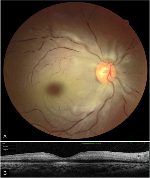
(A) Fundus photography of the right eye showing retinal whitening with a cherry red spot and segmental blood flow consistent with central retinal artery occlusion. (B) Swept source OCT scan showing hyperreflectivity of inner retinal layers

(A) Fundus photography of the right eye showing retinal whitening with a cherry red spot and segmental blood flow consistent with central retinal artery occlusion. (B) Swept source OCT scan showing hyperreflectivity of inner retinal layers