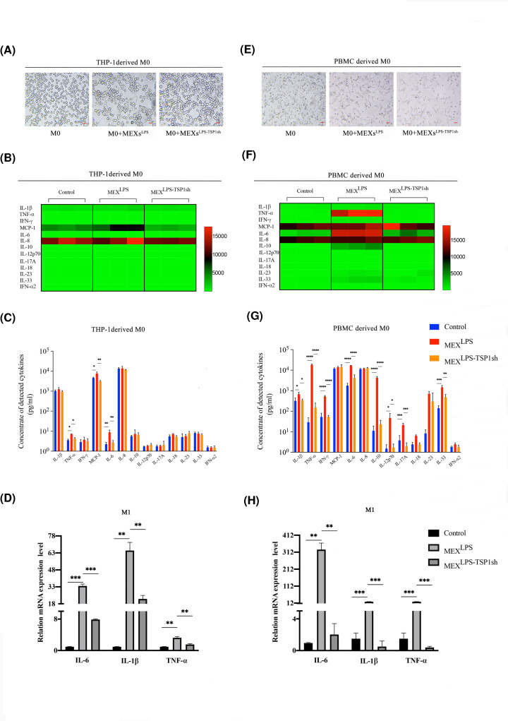
Figure 4. TSP-1 as the main factor in the proinflammatory regulation of macrophages.
(A and E) Morphological changes of THP-1-derived and PBMC-derived macrophages co-cultured with exosomes, bar = 20 μm. (B and F) Heat-map of cytokine expression profiles. THP-1-derived and PBMC-derived macrophages treated with MEXsLPS and MEXsLPS-TSP1sh for 12 h. All samples were run in triplicate. The colors illustrate fold changes (see color scale). Red: up-regulation; green: down-regulation. (C and G) Detection of inflammatory factors levels in culture supernatants of THP-1-derived and PBMC-derived macrophages treated with MEXsLPS and MEXsLPS-TSP1sh for 12 h. (D and H) Expression of IL-6, IL-1β and TNF-α in THP-1-derived and PBMC-derived macrophages when treated with MEXsLPS and MEXsLPS-TSP1sh for 12 h. Data are expressed as the mean ± SD from three experiments, *P<0.05, **P<0.01, ***P<0.001, ****P<0.0001.

