
Figure 1. PIM3 knockout decreased proliferation, viability, and motility in hepatoblastoma cells.
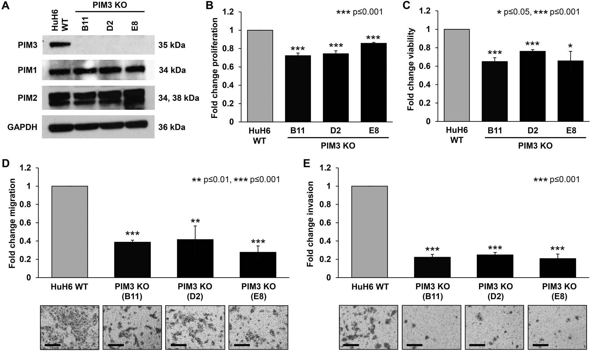
(A) Immunoblotting for PIM kinases in HuH6 wild-type (WT) and PIM3 knockout (KO) cells. PIM3 protein expression was absent as expected in PIM3 KO clones. There was no change in PIM1 and PIM2 protein expression in PIM3 KO cells compared to HuH6 WT cells. GAPDH was used as a loading control. (B) Proliferation and (C) viability were measured using CellTiter 96® and alamarBlue® assays, respectively. All three clones of PIM3 KO cells exhibited significantly decreased (B) proliferation and (C) viability compared to HuH6 WT cells. Data represent at least three biologic replicates and are reported as mean ± standard error of the mean. (D) Migration and (E) invasion were also assessed. HuH6 WT or PIM3 KO cells were seeded into modified Boyden chambers. Inserts were coated on the bottom with collagen which acted as a chemoattractant and a layer of Matrigel™ was added to the top of the insert for invasion. After 24 hours, photographs were taken with representatives shown (panels to the bottom of the graphs) and migration and invasion from at least three biologic replicates reported as mean fold change in number of cells migrating or invading, respectively ± standard error of the mean. Scale bars represent 100 μm. PIM3 KO cells exhibited a significant decrease in (D) migration and (E) invasion compared to HuH6 WT cells.