
Figure 5. PIM3 knockout decreased hepatoblastoma cancer cell stemness.
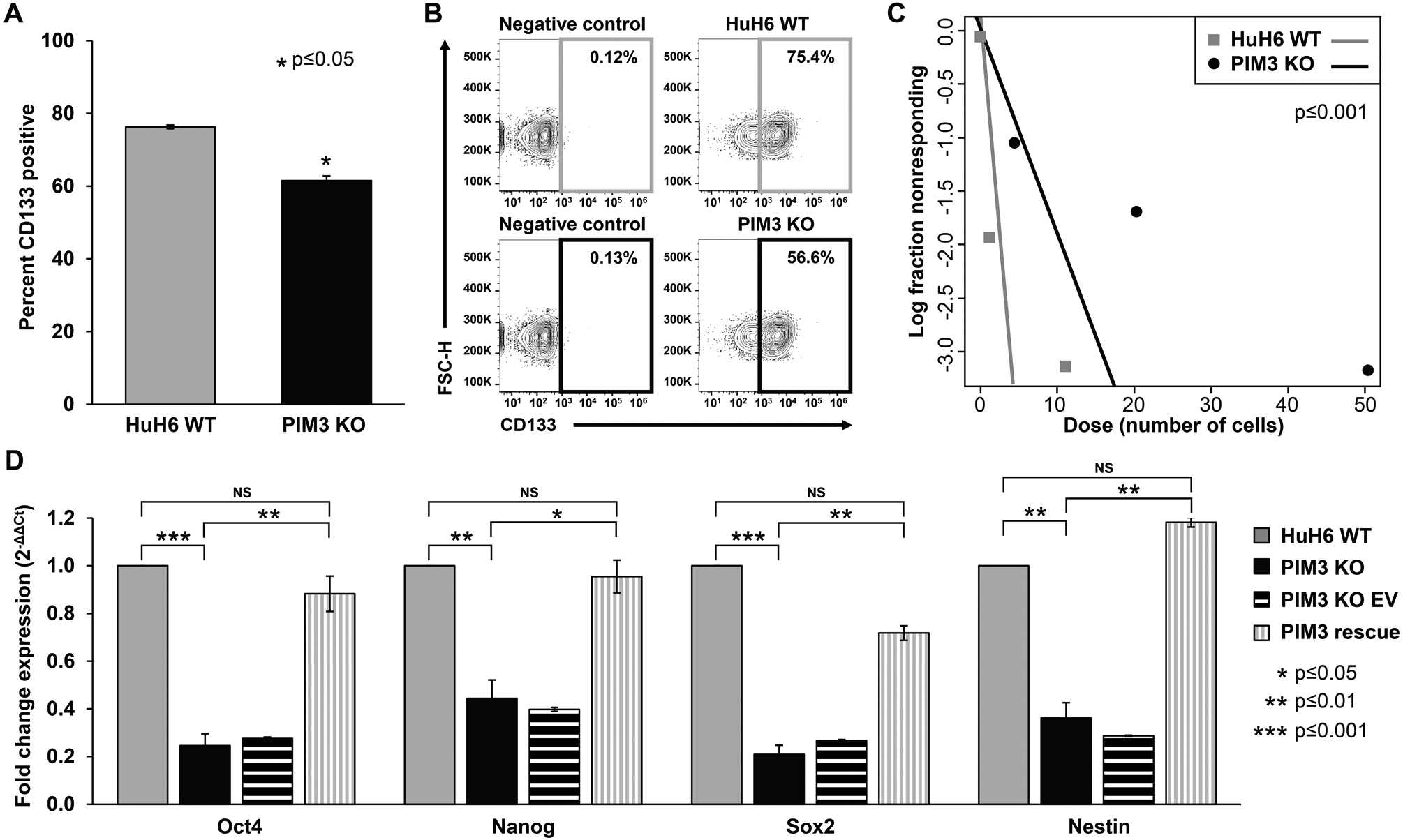
(A) CD133 cell surface expression was determined using flow cytometry. PIM3 knockout (KO) cells had significantly decreased CD133 expression compared to HuH6 wild-type (WT) cells. (B) Representative contour plots with negative staining controls for each cell line are shown. (C) Cells were plated for an extreme limiting dilution assay with decreasing numbers of cells per well. PIM3 KO cells formed spheres less frequently and at higher cell concentrations than HuH6 WT cells, indicating decreased tumorsphere formation ability. (D) Real-time PCR was used to examine the mRNA abundance for Oct4, Nanog, Sox2, and nestin. Gene expression was normalized to β-actin and calculated as fold change to HuH6 WT using the ΔΔCt method. PIM3 KO led to decreased mRNA abundance of these markers compared to HuH6 WT cells, while re-introduction of PIM3 cDNA in PIM3 KO cells led to rescue of mRNA abundance and a return to levels comparable to those seen in HuH6 WT cells. Data represent at least three biologic replicates and are reported as mean ± standard error of the mean. NS: non-significant.