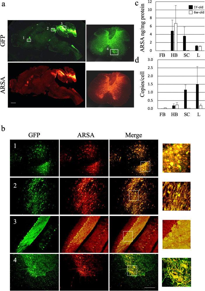
Figure 2.
Expression of GFP and ARSA in the brains of treated MLD mice. AAV9/ARSA (encoding ARSA and GFP) were intrathecally injected via suboccipital puncture into 1-year-old MLD mice. (a) representative images of immunostaining for GFP and ARSA in sagittal brain and spinal cord sections taken 3 months after injection of AAV9/ARSA. Boxes indicate the approximate regions from which the higher magnification images shown in b were acquired. (b) 1, caudate putamen (striatum); 2, inferior colliculus; 3, cerebellum; 4, ventral horn of the cervical spinal cord. Bar: 200 μM. Panels on the right highlight the observed co-localization. (c) ARSA levels in the indicated brain regions measured with ELISAs in MLD mice treated at 6 week (n = 4) and 1 year (n = 5) of age. (d) droplet digital PCR to detect the copy numbers of AAV vector in MLD mice treated at 6 week (n = 4) and 1 year (n = 5) and of age. FB, forebrain; HB, hindbrain; SC, spinal cord; L, liver; ARSA, human arylsulfatase A.

