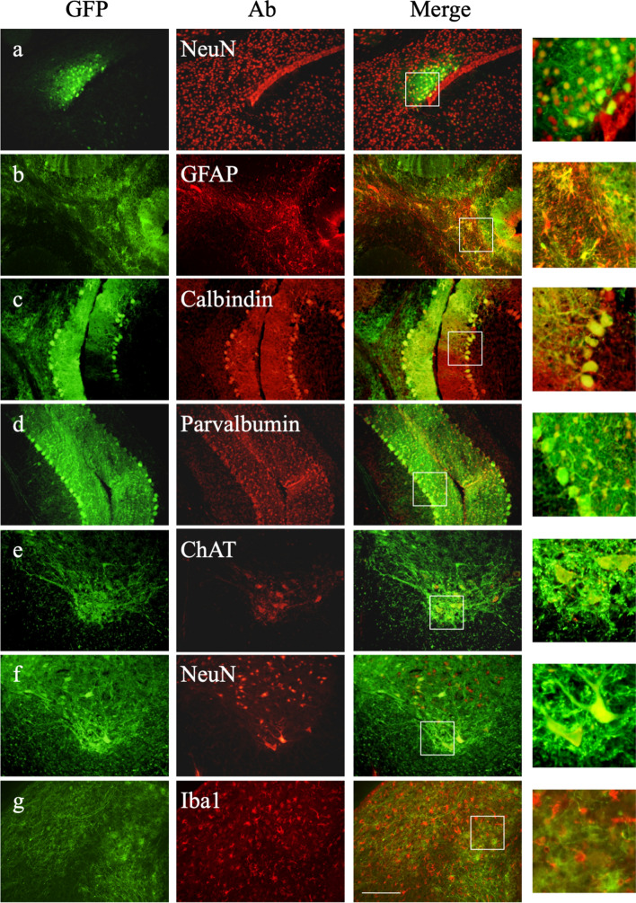
Figure 3.
Immunohistochemical analysis of transduced cells in the brain and spinal cord of treated MLD mice at 1 year of age. Immunostaining for GFP and the neuronal marker NeuN (a), the astrocyte marker GFAP (b), the Purkinje marker calbindin (c), the basket marker parvalbumin in the cerebellum (d), the microglia marker Iba1 (g) in the brain, and ChAT-positive motor neurons (e) and NeuN-positive neurons (f) in the cervical spinal cord, especially the ventral horn. Bar: 200 μM. Panels on the right highlight the observed co-localization.

