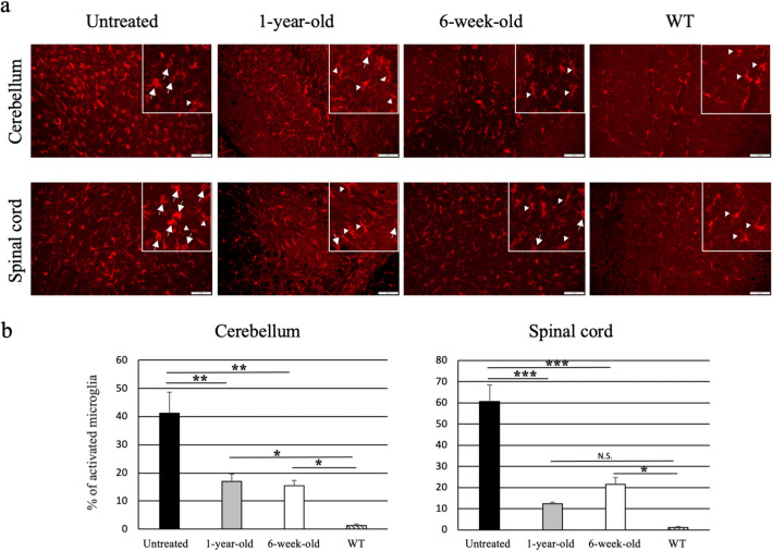
Figure 5.
Reduction of activated microglia in AAV9/ARSA-treated MLD mice. (a) Immunostaining for the microglia marker Iba1 in the cerebellum and spinal cord. (b) Fifteen-month-old untreated mice showed amoeboid microglia cells (arrows), which correspond to activated microglia. MLD mice treated with AAV9/ARSA at 1 year or 6 weeks of age and wild-type (WT) mice showed ramified microglia (arrow heads), which correspond to non-activated microglia. % of activated microglia was calculated as (number of activated microglia/ numbers of Iba1 positive cells). All calculations were done on random three fields of each mouse (untreated: n = 3, 1 year treated: n = 3, 6 week treated: n = 3, and WT: n = 3) Bar: 100 μM. *p < 0.05, **p < 0.01, ***p < 0.0001.

