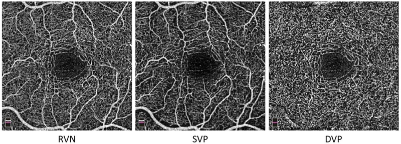
Figure 1. OCTA enface images.
A study subject was scanned at the macula centered on the fovea (i.e., central area with no vessels) with a field of view 3 × 3 mm. Angiographic images (i.e., enface view images) of the vascular slabs, including total retinal vascular network (RVN), superficial vascular plexus (SVP), and deep vascular plexus (DVP), show capillary network around the avascular fovea. Note, there is only the capillary network in the DVP slab, in contrast to RVN and SVP, which mixed capillary network with small vessels (i.e., arterioles and venules).

