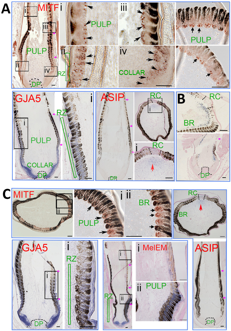
Fig. 5.
Molecular expression during feather formation. Immunostaining and ISH of Partridge Plymouth Rock feather follicles (A), Fayoumi feather follicles (B), and Dark Cornish feather follicles (C). MITF is based on immunostaining. GJA5, melEM, and ASIP are based on ISH. (A) MITF+ cells (red nucleus staining) are present in the basal layer of the feather filament epidermis in longitudinal feather sections (arrows in A, Mitf-i, ii, iii, and iv) and cross-section (Right) of both eumelanin and pheomelanin regions. GJA5 is expressed (blue color) in keratinocytes in collar and ramogenic zones. GJA5 is also expressed in melanocytes in the ramogenic zone in both the eumelanin and pheomelanin zones, but with decreased expression in the more differentiated barb ridges. ASIP is absent in the pulp in longitudinal sections. Cross-sections show that ASIP is weakly expressed in the peripheral pulp adjacent to the rachis region. (B) Fayoumi chicken feathers with autosomal barring pattern (3) are shown for comparison. Both cross and longitudinal sections show lower ASIP expression in the peripheral pulp adjacent to the eumelanin region. (C) MITF immunostaining (Upper) shows positive melanoblasts in both eumelanin and pheomelanin regions in Dark Cornish feather follicles. GJA5 and ASIP expression patterns are similar to the expression patterns shown in Partridge Plymouth Rock feather follicles (A). MelEM (blue nucleus staining) is expressed in melanoblasts in the distal collar and ramogenic zone, with strong expression in the eumelanin region and weak expression in the pheomelanin region. For feather follicle components, please refer to Fig. 6A. BR, barb ridge; DP, dermal papilla; RC, rachis; RZ, ramogenic zone. (A) MITF, GJA5, ASIP panels; (B) Lower panel; and (C) GJA5, MelEM Left panel, and ASIP panels are photomontages in which spliced junctions are indicated by purple arrows. (Scale bars in all panels, 100 μM.)

