Fig. 3.
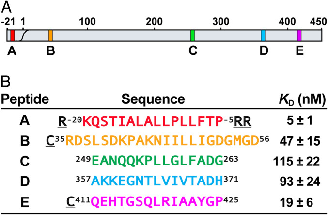
Potential DnaK binding sites on proPhoA and designed peptide models. (A) Schematic of the proPhoA sequence with strong DnaK binding sites identified from a proPhoA peptide array (9) labeled as A, B, C, D, and E and colored in red, orange, green, blue, and magenta, respectively (this color code is used in all figures). (B) Model peptides and their SBD apparent binding affinities (KD) (SI Appendix, Fig. S4). (Underlined residues are not the part of proPhoA sequence.)

