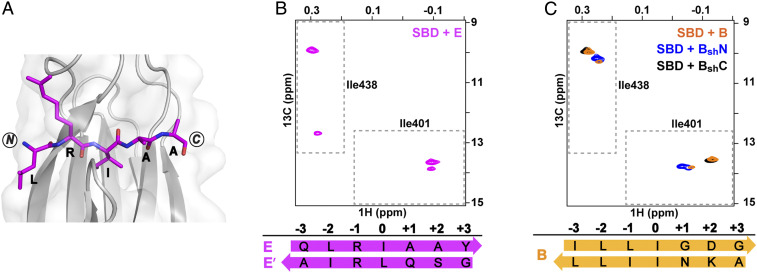
Fig. 5.
Peptides containing proPhoA sites E and B both populate more than one binding mode, one N to C, and another minor state, C to N. (A) Crystal structure of the DnaK SBD (gray) complexed with proPhoA peptide E (magenta). (B) The Ile401 and Ile438 region of 1H-13C-HMQC spectra of ILV-13CH3 ILV-13CH3-DnaK SBD in the presence of unlabeled peptide E (magenta). Two binding modes are populated; the minor C to N binding mode is denoted E’ (SI Appendix, Fig. S8). (C) The Ile401 and Ile438 region of 1H-13C-HMQC spectra of ILV-13CH3 DnaK SBD in the presence of unlabeled peptides B (orange), BshN (blue), and BshC (black). The major resonances for peptide B represent an N to C binding mode with I50 in the central pocket; the minor resonances can be assigned to C to N binding mode with I46 in the 0th pocket (SI Appendix, Fig. S9).

